Welcome to my site
I am a postdoctoral fellow at the Kavli Institute for Systems Neuroscience at NTNU, Trondheim. My long-term goal in science is to unveil the contribution of different types of neurons to brain function
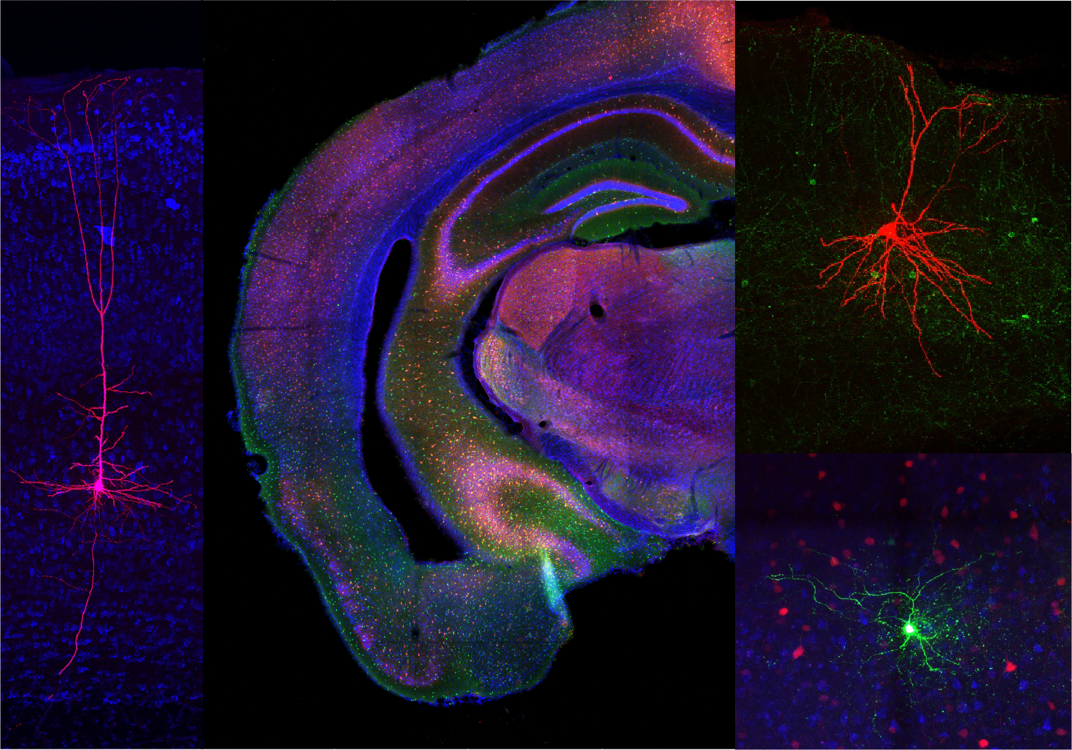
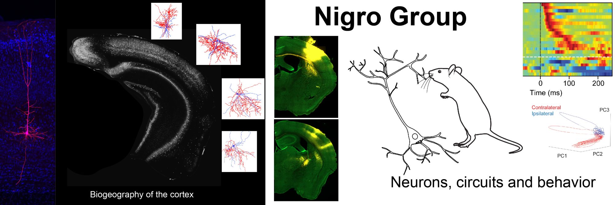
Brain circuits are formed by an astonishing number of different neuron types. Each type of neuron is characterized by specific morphological, physiological and molecular features that allow them to exert specific functions in different circuits. The same features allow neuroscientists to classify these neurons in discrete types, ultimately forming a coherent taxonomy of neurons.
I chose two models to address my research goals: the diversity of cortical GABAergic neurons, and multisensory integration in perirhinal cortex. GABAergic interneurons represent a minority of neurons in the mammalian cortex; however, their wide diversity allows them to participate in specific circuit designs and represent an ideal model to study the relationship between morphology and function. On the other hand, multisensory integration (i.e. the ability to merge information from different senses) is one of the most widespread and distinctive functions of the brain. The perirhinal cortex is a perfect playground to tackle my research interests: it contains a peculiar distribution of GABAergic neuron types that sets it apart from other cortical areas; moreover the perirhinal cortex receives information from all sensory modalities and has been implicated in the creation of multisensory representations of objects.
In this website you will find information about the projects I am working on, including advances, publications, and presentations at conferences. On the Blog page I will post short descriptions of interesting neuroscience articles as well as curiosities of the neuroscience world.